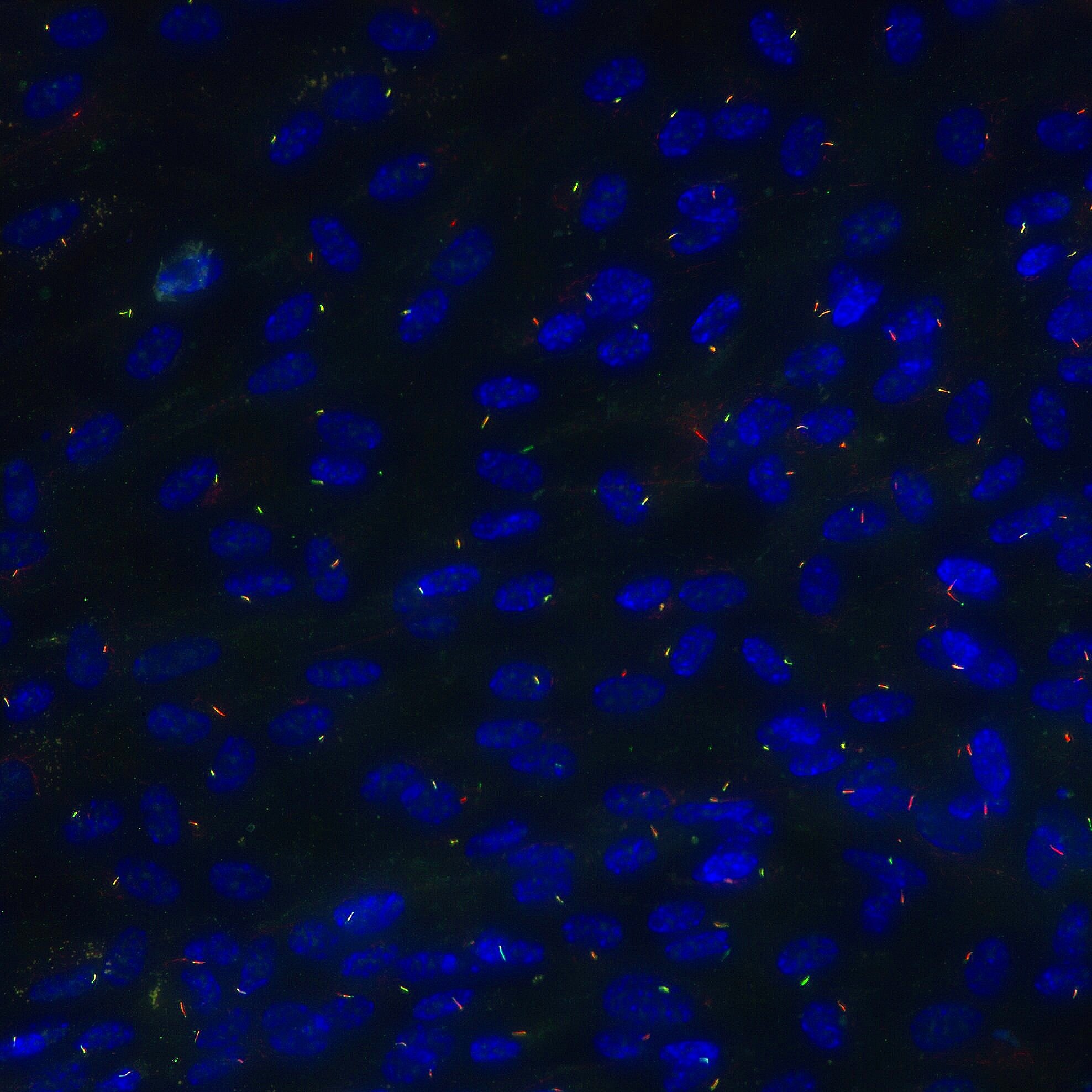
The primary cilium is an antenna-like projection found on nearly every human cell; it extends from the cell body, where it receives and interprets signals, thus allowing cells to respond to their environment. These minuscule projections represent ~1/10,000th of a cell’s volume. Long thought to be vestigial organelles, we now know that loss of cilia is incompatible with human life. Ciliopathies are disorders rooted in ciliary dysfunction and exhibit overlapping clinical features, including developmental delay, intellectual disability, polydactyly, retinal dystrophy, and progressive involvement of the kidney and liver. While individually rare, ciliopathies combined affect 1/500 individuals. Most of our understanding of ciliary biology comes from model systems like the unicellular algae Chlamydomonas reinhardtii, as the structure of these organelles is remarkably conserved. I aim to combine basic science inquiries with questions rooted in determining the pathophysiology of ciliopathies.  

How does the cilium dynamically regulate protein content?

What are the roles of post-translational modifications of the ciliary support structure, the microtubule-based axoneme?

How do ciliopathy-related variants impact these aspects of ciliary function?


Super_res_RPE_slimmer.jpg

A primary cilium as seen on GE DeltaVision OMX Super Resolution Microscope. Acetylated tubulin (red) marks the microtubule-based axoneme and ARL13B (green) labels the membrane.

assistant professor

Department of cell biology

University of Oklahoma health sciences center